A proper assessment does more than confirm a visible patch on the eye: it identifies the lesion, measures its effect on the cornea and vision, and separates irritation that needs symptom relief from growth that needs removal. By the end, you will know what happens during consultation, when observation or non-surgical care is reasonable, what surgery involves, and which questions to ask before choosing a clinic.
Key takeaways
- Expect a slit-lamp examination and questions about irritation, growth and vision changes.
- Observation suits small, stable pterygia without significant corneal or visual effects.
- Surgery evaluation becomes relevant when growth affects vision, comfort or eyelid movement.
- Compare graft techniques, recurrence prevention, recovery time and surgeon experience before deciding.
What happens during a pterygium consultation?
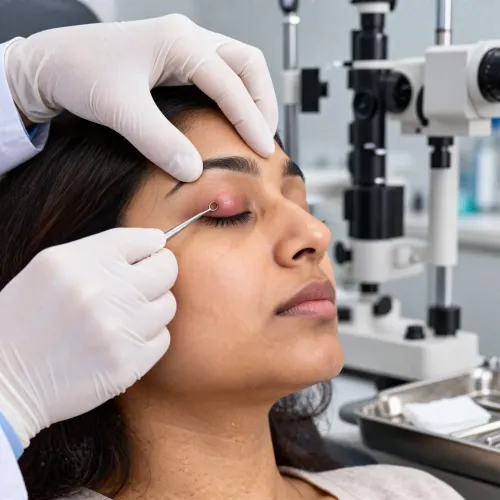
An ophthalmologist confirms a pterygium by examining the growth and checking how it affects vision, rather than judging redness or appearance alone. During a pterygium consultation in Mumbai, expect questions about irritation, dryness, watering, light sensitivity, redness, duration, growth, previous treatment and any change in your spectacle prescription.
The assessment commonly includes:
- Visual-acuity testing for each eye, with and without your current glasses
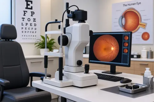
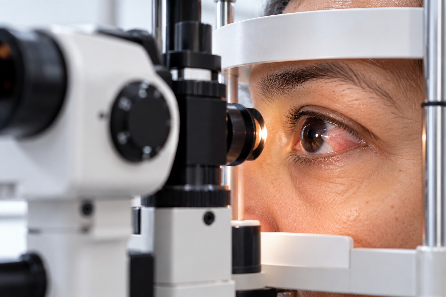
- Slit-lamp examination of the conjunctiva, cornea and limbus
- Measurement of the lesion’s width, thickness and distance from the pupil or visual axis
- Corneal assessment for surface disturbance, scarring and induced astigmatism
- Review for inflammation, discharge or other signs of ocular-surface disease
A pinguecula is a raised yellowish conjunctival spot that does not grow onto the cornea. A pterygium extends across the limbus onto the cornea, often with a triangular wing-shaped appearance.
The doctor also checks whether redness represents infection, allergy, chronic inflammation, a conjunctival tumour or another limbal lesion; an unusual, rapidly changing or heavily vascular growth may need additional investigation.
The consultation should connect measurements to function. A lesion close to the pupil can threaten the visual axis, while corneal distortion can blur vision or change your spectacle power before the growth reaches the pupil. Documented progression, reduced acuity or persistent symptoms gives more useful treatment information than cosmetic prominence alone.
How do examination findings determine observation or non-surgical care?
A small, thin growth that remains stable and sits away from the pupil usually calls for observation rather than removal. The ophthalmologist documents its location, size and thickness at the slit lamp, checks visual acuity, and looks for corneal distortion or a change in spectacle power.
A growth nearing the visual axis deserves closer follow-up even before it affects sight.
The examination findings that support monitoring or non-surgical care include:
- Stable size and a clear gap between the growth and the pupil: schedule repeat examinations to detect progression.
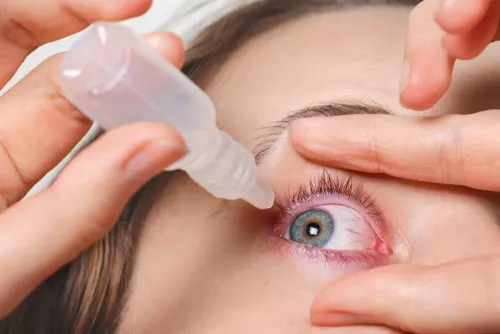
- Mild redness or irritation without corneal distortion: use preservative-free lubricating drops and reduce wind, dust and smoke exposure.
- Recurrent surface irritation with visible inflammation: treat the inflammation with prescribed eye drops; do not use steroid drops without an ophthalmologist’s direction.
- A thick or broad lesion that remains outside the visual axis: monitor its measurements and symptoms rather than judging treatment by appearance alone.
Sunglasses that block ultraviolet light, a brimmed hat and limiting prolonged sun exposure can reduce environmental irritation. Drops and protection relieve symptoms; they do not remove an established growth. If it enlarges toward the pupil, causes induced astigmatism, reduces vision or continues inflaming despite treatment, the follow-up plan changes to a surgical discussion.
These are central pterygium treatment details in Mumbai and elsewhere: function and progression matter more than redness or cosmetic prominence alone.
When does a pterygium surgery evaluation become appropriate?
Removal becomes appropriate when the pterygium causes ongoing symptoms or begins to affect corneal shape and sight, not merely because the eye looks red. Consider a pterygium surgery evaluation in Mumbai if you have:
- Persistent dryness, grittiness, burning or foreign-body sensation despite lubricating drops and treatment of surface inflammation.
- Recurrent episodes of redness and inflammation.
- Corneal distortion that produces irregular astigmatism.
- A changing spectacle prescription, especially when one eye’s power changes without another clear cause.
- Reduced visual acuity or blur that interferes with reading, driving or work.
- Growth that is advancing toward the pupil or visual axis.
- Substantial cosmetic concern after the doctor has discussed recurrence and surgical risks.
A growth near the visual axis deserves prompt assessment because further extension can obstruct vision and make removal more consequential. Do not wait for it to cover the pupil; documented progression and induced astigmatism are important reasons to discuss surgery earlier.
The evaluation should compare vision and refraction with earlier records and examine whether the blur comes from corneal distortion rather than the tissue alone. For a large lesion, a growth encroaching on the cornea, or suspected irregular astigmatism, ask whether corneal topography is needed.
The surgeon should also confirm that the lesion is a pterygium rather than a pinguecula, tumour or another limbal condition before recommending removal.
Which surgical and practical treatment options should you compare?
Most pterygium removal is outpatient surgery under local anaesthesia, but confirm the setting and sedation plan. Age, anxiety, general health, lesion complexity and facility equipment can change that plan. Ask how astigmatism and vision will be measured before and after surgery.
| Option | Practical comparison | When to discuss |
|---|---|---|
| Excision with conjunctival autograft | The removed tissue is covered with your own conjunctiva. Sutures are widely used; fibrin glue can shorten surgery and reduce suture discomfort, but adds cost and handling-related risks. | Common primary cases; compare graft size, technique and surgeon experience. |
| Amniotic membrane transplantation | Preserves conjunctiva and covers a larger surface, but is not automatically better than an autograft. | Large defects, recurrent or complex disease, or when conjunctiva must be preserved for future glaucoma surgery. |
| Mitomycin C | An adjunct that can reduce fibrovascular regrowth, but requires carefully selected use because excessive exposure can damage the ocular surface. | Ask why it is needed, the concentration and exposure protocol, and how safety will be monitored. |
When comparing pterygium treatment options in Mumbai, request a written estimate that identifies:
- Preoperative examination, refraction or corneal topography, surgeon and operating-facility charges
- Graft material, fibrin glue or mitomycin C, if used
- Anaesthesia, prescribed drops and each postoperative review
- Management of recurrence, infection, graft problems or other complications
Do not treat a quoted total as universal: technique and facility change the final amount. Ask whether the surgeon has verifiable ophthalmology registration, corneal or ocular-surface training, and substantial experience with the proposed graft method.
What should you ask about recovery, recurrence and surgeon experience?
Postoperative drops are part of the treatment, not an optional extra. Ask how often to use the antibiotic, steroid and lubricating drops, when each will stop, and whether you must taper the steroid. Contact the surgeon promptly for worsening pain, increasing redness or swelling, pus-like discharge, sudden vision loss or severe light sensitivity.
Protect the eye after healing as well as during recovery. Wear sunglasses that block 99–100% of UVA and UVB, use a hat outdoors, and reduce direct ultraviolet exposure; recurrence can appear months or years later.
Surgery lowers the risk but does not eliminate it, particularly with young age, heavy UV exposure, persistent inflammation, a large fleshy growth or previous recurrence.
“Pterygium specialist in Mumbai” is a search label, not a standard qualification. Check the ophthalmologist’s registration, ophthalmic surgical training and experience with corneal or ocular-surface procedures. Recurrent disease deserves specific discussion because scarring and limited conjunctival tissue can make grafting more difficult.
Ask these questions before consenting:
- Which graft method will you use, and why is it suitable for my eye?
- How will you measure vision and astigmatism before and after surgery?
- How many pterygium procedures do you perform, and how do you manage recurrence or graft displacement?
- What will my drops, reviews and warning-sign instructions include?
- Does the written estimate include examination, measurements, surgeon and facility fees, graft or glue, drops and complication care?
Dr Nusrat Bukhari's Eye Clinic can provide these details in a written treatment plan, allowing you to compare technique, follow-up and total scope rather than the excision fee alone.
Related service
Pterygium Management A pterygium—commonly known as “surfer’s eye”—is a fleshy growth on the white part of the eye that can cause redness, irritation, and blurred vision... View service → |
Frequently asked questions
What happens during a pterygium consultation in Mumbai?
An ophthalmologist examines the growth with a slit lamp, checks the cornea and tear film, measures vision and asks about irritation, dryness, watering, light sensitivity, redness, growth and prescription changes.
How do examination findings determine observation or non-surgical care?
Observation is appropriate when the pterygium is small, stable and does not affect vision or corneal shape. Lubricating eye drops, sunglasses and protection from dust and ultraviolet light can help manage irritation.
When is a pterygium surgery evaluation appropriate?
Arrange a surgery evaluation when the growth causes blurred or distorted vision, recurrent inflammation, persistent discomfort, restricted eyelid movement or documented progression toward the visual axis.
Which pterygium treatment options should you compare?
Ask about observation, symptom control and excision with conjunctival autograft. Compare the graft method, sutures or tissue adhesive, recurrence prevention, recovery restrictions and follow-up schedule.
What should you ask about recovery, recurrence and surgeon experience?
Ask how long redness and discomfort usually last, when you can work or exercise, which warning signs need urgent review, how recurrence is monitored and how frequently the surgeon performs pterygium surgery.
Keywords